LIGHT COHERENCE
Is this Property Important for Photomedicine?
Tiina I. Karu
Institute of Laser and Information Technologies, Russian Academy of Sciences, Troitsk 142190, Moscow Region, Russian Federation
tkaru@isan.troitsk.ru
Historically, one of the most topical and widely discussed issues by the laser phototherapy ("laser biostimulation") clinical community has been the question of whether the coherence of laser radiation has additional benefits from a therapeutical point of view, as compared with monochromatic or quasimonochromatic light from a conventional light source (i.e., properly filtered light from a lamp) or LED (light-emitting diode) with the same wavelength and intensity as a laser beam. Nowadays, the problem of importance of light coherence in biological experiments is not as hot a topic as it used to be 10-20 years ago. First, the mechanism of "laser biostimulation" is now known to be a photobiological process (Karu, 1986; 1987) due to the absorption of light by cellular photoacceptors, cytochrome c oxidase in particular (Karu, 1999, 2007). Second, LED’s emitting noncoherent light are now used widely and successfully in clinical practice. The problem concerning light coherence, however, is once more dual. First, this problem arises from time-to time in spite of the successful use of LED’s in clinical practice. Second, it is possible that in deeper tissue layers, in addition to the photobiological effects, there may be beneficial effects of coherent (laser) light.
Two aspects of the topic of the present paper must be distinguished: the coherence of lights itself, and the coherence of the interaction of light with matter (i.e., biomolecules, tissues).
Coherence of Light
Coherence is one of the unique properties of laser radiation. It arises from the stimulated light emission process, providing the amplification of emitted photons, which have a definite relation to each other, i.e., they are in phase. The coherent properties of light are described by temporal and spatial coherence. Temporal coherence is characterized by coherence length, Lcoh. Lcoh expresses the propagation distance over which coherence of the beam is kept. Temporal coherence also tells us how monochromatic a source is. The temporal coherence of light is determined by the spectral width,
 , since the coherence time,
  coh, during which light oscillates at the point of irradiation has a regular and strongly periodic character:
(1.1)

Here
is the spectral width (full width at half maximum) of the beam in Hz. Since light propagates at the rate c=3×1010 cm/sec, the light oscillations are matched by the phase (i.e., they are coherent) on the length of light propagation, Lcoh, (i.e., the measure of temporal or longitudinal coherence):
(1.2)
The more monochromatic is the light, the longer is the length where the light field is coherent in volume. For a single mode (single frequency) He-Ne laser (λ = 632.8 nm), Lcoh>>1 m. But for an LED emitting at λ = 800 nm (12,500 cm-1),
= 160 cm-1 (or Δλ = 10 nm), and Lcoh = 1/160 cm-1 ≅ 60 μm. For a multimode (multi-frequency) He-Ne laser with = 500 MHz, Lcoh = 60 cm.
The spatial or lateral coherence of the laser beam describes the correlation between the phases of the light field in a lateral direction. The size of the spatial coherence,
 coh
, is connected with the divergence (ϕ) of the light beam at the point of irradiation:
coh
, is connected with the divergence (ϕ) of the light beam at the point of irradiation:
(1.3)

With conventional light sources, the size of the emitting area is significantly larger than the light wavelength, and various parts of this area emit light independently or noncoherently. In this case, the size of the lateral coherence,
coh, is significantly less than the diameter of the light beam.
Coherence of Light Interaction with Biomolecules, Cells, and Tissues
The coherent properties of laser light can not be maintained when the beam interacts with a biotissue at the molecular level. This conclusion was made first in the paper by Karu (1987). Under physiological conditions, the absorption of low-intensity light by biological systems is purely of noncoherent nature (i.e., photobiological), because the rate of decoherence of photoexcitation (the processes that limit the appearance of quantum effects, and turns them into classical phenomena) is many orders of magnitude higher than the rate of photoexcitation. The time of decoherence of photoexcitation is the factor that determines the interaction of light quanta with surrounding molecules. Under normal conditions (at room temperature, and low light intensities used in "laser biostimulation"), the time of decoherence is less that 10-12 sec.
Recall that the average excitation time depends on the light intensity. For example, at an intensity of 1 mW/cm2, this time is around 1 sec. At 300° K (room temperature) in condensed matter for compounds absorbing monochromatic visible light, the light intensity at which the specific interactions between coherent light and matter start to occur was estimated to be above the GW/cm2 level (Karu, 1987). Note that the light intensities used in clinical practice of "laser biostimulation" are not higher than tens or hundreds of mW/cm2. Indeed, the stimulative action of various bands of visible light at the level of organisms and cells was known long before the advent of the laser (for a review, see: Karu, 1987, 2007). Also, specially designed experiments at the cellular level have provided evidence that coherent and noncoherent light with the same wavelength, intensity, and irradiation time provide the same biological effect (Karu et al., 1982a, b, 1983; Bertoloni et al., 1993). The successful use of LED’s (light-emitting diodes) in many areas of clinical practice over the last decade also confirms this conclusion.
Therefore, it is possible that the effects of light coherence are manifested at the macroscopic (e.g., tissue) level at various depths (L) of irradiated matter. Figure 1 presents the coherence volumes (Vcoh) and coherence lengths (Lcoh) for four different light sources. Recall that the coherence length (Lcoh) is the propagation distance from a coherent light source to a point where electromagnetic wave maintains a specified degree of coherence. Coherence volume (Vcoh) is coherence length (ΔLcoh) multiplied to coherence area. Figure 1A presents the data for two coherent-light sources (He-Ne and semiconductor (diode) lasers as typical examples of therapeutic devices). Let us mention that the divergence of the beam of the semiconductor laser tends to be larger (compare two examples in Figure 1A), which is due to the specificity of the non-Gaussian beam geometry. A Gaussian beam is a beam of electromagnetic radiation whose transverse electric (TE) field and intensity distributions are well approximated by Gaussian function. In this case, the laser is operating in TEM00 mode (single mode) of its optical resonator.
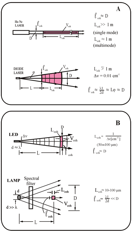
Figure 1. Coherence volumes and coherence lengths of light from: (A) laser and (B) conventional sources when a tissue is irradiated. Lcoh, length of temporal (longitudinal) coherence;
Figure 1B presents the respective data for noncoherent light (LED and spectrally filtered light from a lamp). Comparing Figures 1A and B shows that large volumes of a tissue are irradiated only by laser sources with monochromatic radiation. For noncoherent-radiation sources (Figure 1B), the length of the coherence, Lcoh, is small. This means that only surface layers of an irradiated substance can be achieved by noncoherent light from LED’s and conventional sources. The numeral value of the thickness of these surface layers depends on the irradiation parameters (wavelength and intensity), as well as on tissue characteristics, and can range from millimeters to centimeters.
The spatial (lateral) coherence of the light source,
coh, is unimportant, due to the strong scattering of light in biotissue when propagated to the depth L>>SC, where SC is the free pathway of light in relation to scattering. This is because every region in a scattering medium is illuminated by radiation with a wide angle (ϕ ≈1 rd). This means thatcoh ≅ λ, i.e., the size of spatial coherence, coh, decreases to the wavelength of light used for irradiation (Figure 1). One has to note that there are differences between conventional laser (e.g., He-Ne laser) and semiconductor laser beam properties. In semiconductor lasers, which are also used for therapeutical purposes, the beam is not Gaussian and the beam divergence tends to be larger.
Thus, the length of longitudinal coherence (Lcoh) is important when bulk tissue is irradiated, because this parameter determines the volume of the irradiated tissue, Vcoh. In this volume, the random interference of scattered light waves and the formation of random nonhomogenities of intensity in space (speckles) occur. For noncoherent-light sources, the coherence length is small (tens to hundreds of microns). For laser sources, this parameter is much higher. Thus, the additional therapeutic effect of coherent radiation, if this indeed exists, depends not only on the length of Lcoh, but also, and even mainly, on the penetration depth into the tissue due to absorption and scattering, i.e., by the depth of attenuation. Table 1 summarizes the qualitative characteristics of the coherence of various light sources, as discussed above.
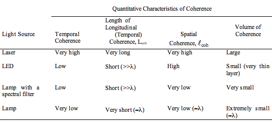
Table 1. Comparison of coherence (temporal and spatial) of various light sources used in clinical practice and experimental work (Karu, 2003).
The difference in the coherence length, Lcoh, is unimportant when thin layers of biological objects are irradiated, inasmuch as the longitudinal size of the irradiated object, Δ
, is less than Lcoh for any source of monochromatic light (i.e., filtered lamp light, LED, laser). Examples are a monolayer of cells, or an optically thin layer of cells in suspension (Figures 2A, B). Recall that layers of cells with a small optical depth, through which light passes easily, because the absorbers do not shadow each other, is considered to be optically thin. Indeed, experimental results (Karu et al., 1982a, b; 1983; Bertoloni et al., 1993) on these models provided clear evidence that the biological responses to coherent and noncoherent light with the same parameters are equal in these two model systems.
Speckles
The situation is quite different when a bulk tissue is irradiated (Figure 2C). The coherence length, Lcoh, is very short for noncoherent-light sources, and can play some role only on surface layers of the tissue with thickness Δ
surface. For coherent-light sources, the coherence of the radiation is retained along the entire penetration depth, L. The random interference of light waves of various directions occurs over this entire distance in bulk tissue (Δbulk). As a result, a speckle pattern of intensity appears. A speckle is a mottled pattern that arises when laser light falls on a non-specular reflecting surface. Speckle patterns occur because of the interference of a large number of elementary waves that arises when coherent light is reflected from a rough surface, or when coherent light passes through a scattering medium. Random interference creates a three-dimensional random intensity distribution of light. Maximal values of the intensity appear at the random constructive interference. The minima (i.e., regions of zero intensity) occur at the random destructive interference. The dimensions of these speckles at every occurrence of directed random interference are approximately within the range of the light wavelength, λ. The coherent effects (speckles) appear only at the depth, Lcoh.
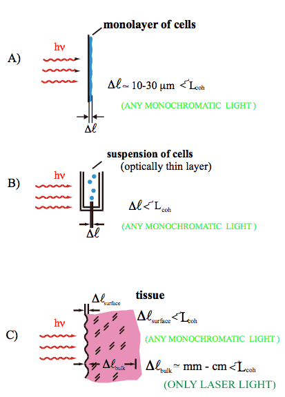
Figure 2. Depth (Δ
These laser-specific speckles cause a spatially nonhomogeneous deposition of light energy, and lead to statistically nonhomogeneous photochemical processes, an increase in temperature, changes in local pressure, deformation of cellular membranes, etc. Note that the same coherent light source may exhibit observable interference effects in some situations and not in others. Also, a non-coherent source exhibits random phase patterns both temporally and spatially, but this radiation is not capable of exhibiting stable or observable interference effects. For example, an incandescent light beam or a flashlight beam reflected on a surface exhibits no speckle patterns (Mobley, J. and Vo-Dinh, T., 2003).
For nonpolarized coherent light, the random speckles are less pronounced (they have lower contrast), as compared to the speckles caused by coherent polarized light. A special feature of nonpolarized coherent radiation is that the regions with zero intensity appear less often, as compared with the action of coherent polarized light. Thus, the polarization of laser light causes brighter random intensity gradients, which can enhance the effects of light coherence when tissue is irradiated.
In scattering biotissue, the main role is played by the coherence length of the beam, inasmuch as this parameter determines the depth of tissue where the coherent properties of the light beam can potentially be manifested, depending on the attenuation. This is the spatial (lateral) coherence of the beam, i.e., its directivity, which plays the main role in the delivery of light into biotissue. Recent experiments (Fixter et al., 2011) showed that the spatial coherence of laser light (λ = 532 nm) was not lost when the light went through a static tissue, but it was partially lost when there was a flow of fluid through the tissue. The volumetric flow rate was directly correlated to the loss of spatial coherence length. Higher flow rate produced a shorter coherence length. In addition, the direction and orientation of laser radiation could be important factors for some types of tissues (e.g., dental tissue) that have fiber-type structures (filaments). In this case, waveguide propagation effects of light can appear, those providing an enhancement of penetration depth. Indeed, the experimental data suggest that the coherence length can play a role in laser phototherapy of gingival inflammation (Qadri et al., 2007).
Concluding Remarks
The qualitative picture described above explains why coherent and noncoherent light with the same parameters (i.e., wavelength, dose, intensity) produce the same biological effects on cell monolayers (Karu et al., 1982a, b), and in dilute cell suspensions (Karu et al., 1983; Bertoloni et al., 1993), as well as on tissue surfaces (e.g., the healing of peptic ulcers; Karu et al., 1984; Sazonov et al., 1985). In these cases, the healing effect of irradiation is occurring via absorption of light by photoacceptors (cytochrome c oxidase in particular, Karu, 1999, 2003). However, some additional (therapeutic) effects from coherent and polarized radiation, in addition to those caused by light absorption by photoacceptors molecules, can appear in deeper layers of bulk tissue.
A theoretical consideration was published by Rubinov and Afanas’ev in 2005. They considered, first, the gradient effects arising upon interaction of a biological system with spatially inhomogeneous radiation (a mechanism that may be characteristic for coherent radiation). Secondly, they considered possible light-induced dipole-dipole interactions between biological particles (a mechanism that probably might occur by irradiation both with coherent and noncoherent radiation). To date, no experimental work has been performed to study these possible additional effects qualitatively and quantitatively. In any case, the main therapeutic effects occur due to light absorption by cellular photoacceptors.
References
Bertoloni, G., Sacchetto, R., Baro, E., Ceccherelli, F. and Jori, G. (1993). Biochemical and morphological changes in Echerichia coli irradiated by coherent and noncoherent 632.8 nm light. J. Photochem. Photobiol. B: Biol. 18, 191–196.
Fixter, D., Hamootal, D., Ankri, R. and Zalevsky, Z. (2011). Determination of coherence length in biological tissues. Lasers Surg. Med. 43, 339-342.
Karu, T. (1986). Biological action of low-intensity visible monochromatic light and some of its medical applications. In: Laser. Ed. by G.Galletti, Bologna: Monduzzi Editore, pp. 25–29.
Karu T.I. (1987). Photobiological fundamentals of low-power laser therapy. IEEE J. Quantum Electron. QE-23, 1703–1717.
Karu T. (1999). Primary and secondary mechanisms of action of visible-to-near IR radiation on cells. J. Photochem. Photobiol. B: Biology, 49(1), 1-17.
Karu, T.I. (2003). Low power laser therapy. In: Biomedical Photonics Handbook. Ed. by Tuan Vo-Dinh, Boca Raton: CRC Press, Ch. 48, pp. 48-1–48-25.
Karu, T.I. (2007). Ten Lectures on Basic Science of Laser Phototherapy. Prima Books AB, Grängesberg (Sweden) 2007.
Karu, T.I., Kalendo, G.S., Letokhov V.S. and Lobko, V.V. (1982a). Dependence of biological action of low-intensity visible light upon HeLa cells on irradiation parameters: coherence, dose, wavelength and irradiation mode. Sov. J. Quantum Electron. 12, 1134–1138.
Karu, T.I., Kalendo, G.S., Letokhov V.S. and Lobko, V.V. (1982b) Biostimulation of HeLa cells by low intensity visible light. Nuovo Cimento D, 1, 828–840.
Karu, T.I., Tiphlova, O.A., Letokhov V.S. and Lobko, V.V. (1983). Stimulation of E. coli growth by laser and incoherent red light. Nuovo Cimento D, 2, 1138–1144.
Karu, T.I., Letokhov, V.S., Lobko, V.V. Novikov, V.F. and Paramonov, L.V. (1984). Phototherapy of gastric and duodental peptic ulcer patients based on cell stimulation with low-intensity red light. Vopr. Kurortol. Fizioter. Lech. Fiz. Kult. (Moscow), №1, 36–39.
Mobley, J. and Vo-Dinh, T. (2003). Optical properties of tissue. In: Biomedical Photonics Handbook. Ed. by Tuan Vo-Dinh, Boca Raton: CRC Press, Ch. 2, pp. 1-75.
Qadri, T., Bohdanecka, P., Tunér, J., Miranda, L., Altamash, M. and Gustafsson, A. (2007). The importance of coherence length in laser phototherapy of gingival inflammation–a pilot study. Lasers Med. Sci., DOI 10.1007/s10103-006-0439-1
Rubinov, A.N. and Afanas’ev, A.A. (2005). Nonresonance mechanisms of biological effects of coherent and incoherent light. Optics Spectrosc., 98, 943–948.
Sazonov, A.M., Romanov, G.A., Portnoi, L.M., Odinokova, V.A., Karu, T.I., Lobko, V.V. and Letokhov, V.S. (1985). Low-intensity noncoherent red light in the complex treatment of peptic ulcers. Sov. Med., №12, 42–45.
06/04/11