BIOLUMINESCENCE LABORATORY
John Lee
Department of Biochemistry and Molecular Biology
University of Georgia, Athens, GA 30602
jlee35uga@gmail.com
These laboratory exercises are designed for students of high-school and college. The aim is to demonstrate several properties of bioluminescence systems, both in organisms and extracts in solution. Access to laboratory facilities is desirable, but not necessary for most of the projects. The aim of the experiments is to demonstrate several features that are common to biological systems.
1. There are a variety of luminous organisms, each having a different chemistry.
2. The bioluminescence system can be extracted into solution, and is shown to be the result of an enzyme-catalyzed chemical reaction, i.e., it is not dependent on the "living state".
3. The light emission depends on air (oxygen).
4. There is a range of bioluminescence colors, and conditions can be discovered to "tune" the color.
5. Bioluminescence reactions have important applications for ultra-sensitive analyses.
There are a great variety of bioluminescent organisms to be discovered in Nature, but for these laboratory demonstrations, it is sufficient to just concentrate on the firefly and bioluminescent marine bacteria.
The 10-minute dark-adapted eye is in fact a very sensitive light detector, but for quantitative measurements, a photoelectric device is needed. In research labs a special instrument called a "luminometer" is often used, but any instrument that uses a photo-detector can be modified, such as a fluorometer or an absorption spectrometer with the light-source turned off, or a scintillation counter operated in out-of-coincidence mode. Care must be taken to exclude room light from the sample during measurement. A piece of heavy black cloth works well.
FIREFLY BIOLUMINESCENCE
Preparations
Firefly Buffer. Dissolve 0.8 g of glycine and 0.25 g of ammonium bicarbonate in 200 ml of distilled water. Adjust the pH to about 7.6 with dilute HCl or NaOH.
ATP Solution. Dissolve 0.1 g of adenosine triphosphate (ATP) and 0.05 g MgCl2 in 100 ml of firefly buffer.
1. Extract the bioluminescence system in vitro. Fireflies are common during the Summer months in the Americas and Asia. If they are so available, then collect 10-20 specimens (a butterfly net works well) and allow them to dry thoroughly by whatever means available, freeze-dry, desiccate, but avoid any heat. Cut off the tails where the yellow light organ is obvious, and make a "cold extract" of the tails by grinding with about 1 g of clean sand in 3 mL of cold firefly buffer. A mortar and pestle is handy for this, and keep everything cold by working in a cold room or by surrounding the mortar with ice. Pipette the extract into a handy container (e.g., test tube) and keep cold. Discard the particulate left-over by filtration or by using a lab centrifuge. If collecting insects in the field is not practical, dried firefly tails may be purchased from several laboratory supply companies. Keep the cold extract ice cold.
Now make a "hot extract" by repeating the grinding with fresh tails in hot, not boiling, firefly buffer. Cool the test tube containing the extract again in ice.
2. To "see" an enzyme substrate reaction at work. In the dark room measure out about 1 mL of cold extract, and allow this and the hot extract to come to room temperature (RT), or nearly so to your touch. You will notice a low level of yellow light coming from the RT-cold extract sample, which will soon die away. Add the RT-hot extract quickly (with a syringe or Pasteur pipette) to the 1 mL RT-cold extract, and you will get a flash of light. This experiment is an example of a luciferase (enzyme) luciferin (substrate) reaction first reported and named in the late 19th Century by R. DuBois, but in 1947, McElroy showed that the hot extract was in fact ATP, not firefly luciferin.
3. Assay of adenosine triphosphate. Take another 1 mL of cold extract and bring to room temperature, and squirt in 1 mL of ATP solution, and you will see the same flash of yellow light.
4. Bioluminescence color shift. To the final 1 mL of room temperature cold extract, add several drops of 10% acetic acid. Then immediately add another 1 mL of ATP. The light will now be a dull red. It is hard to get the amount of acetic acid added to give just the right acidity, so if this does not work, instead of the acetic acid, add 1 drop of 10% zinc chloride, mix well, followed by the ATP. This will also produce the red bioluminescence.
See also: Firefly Kits
BACTERIAL BIOLUMINESCENCE
It is relatively easy to culture bioluminescent marine bacteria from a natural source, and this gives experience in standard microbiolgical techniques. After growing them on agar plates or in liquid medium, it will be shown how the light is dimmed or extinguished on removal of oxygen (air). For preparation of the bacterial luciferase, some access to a biochemical laboratory is required, as there is no easily located commercial source.
Preparations.
Microbiological techniques. Refer to any undergraduate laboratory manual such as “Sourcebook of Experiments for the Teaching of Microbiology” (S.B.Primrose and A.C. Warlaw, eds.). Academic Press, 1983.
Bacterial Buffer. In 100 ml of distilled water, dissolve 0.25 g of dibasic sodium phosphate and 0.21 g of monobasic potassium phosphate. Adjust the pH to about 7.2. This is a standard Na/K buffer, and should have a pH of 7.2. Be careful to note that these phosphate salts come with several different basicities and hydrations, so if your salts have different formulae from the above, you need to adjust the weights so that the final phosphate molarity is 50 mM.
Bacterial Nutrient Medium. In 100 ml distilled water, dissolve 2.5 g NaCl, 0.5 g tribasic sodium phosphate, 0.2 g monobasic potassium phosphate, 0.05 g of dibasic ammonium phosphate, 0.01 g MgSO4 , 1 g of Bactopeptone, 0.3 ml of glycerol, and 1 g of Bacto-agar. Carefully bring this to the boil, and while still hot, dispense about 5 ml into each of about 20 Petri plates. Replace each lid, then steam sterilize all 20 plates. Wait until the plates are cool to room temperature before spreading any bacteria onto the agar surface.
To prepare the liquid medium for cell culture and luciferase preparation, start with 500 mL of distilled water, and add the other ingredients in five times amounts, except leave out the Bacto-agar. The flask with a completely dissolved solution needs to be fitted with a cotton stopper, and steam sterilized according to any microbiological manual. When cooled to room temperature it is ready for inoculation.
NADH Solution. In 10 ml of bacterial buffer, dissolve 0.1 g of NADH (reduced nicotine adenine dinucleotide). Store refrigerated.
FMN Solution. In 1 liter of water dissolve 0.05 g of FMN (flavin mononucleotide). Store refrigerated.
Dodecanal Suspension. To 10 ml bacterial buffer, add 1 drop of a dodecanal (or decanal) suspension in 1 mL methanol, and shake vigorously for 1 minute. This will yield a cloudy suspension. For the reaction, this needs to be used less than 2 hours after preparation.
1. "Capture" the wild bacteria. Bioluminescent bacteria can be cultivated from a sample of sea water by spreading a few tenths of a milliliter onto Petri plates containing the salt water nutrient agar prepared as above. Sterile wire loop techniques must be used. Luminous blue spots from single colonies will appear within 48 hours at RT. A more successful method is to allow a squid or fish to putrify for a week or so under a fume hood (of course!). In the dark, luminous spots from bacteria will be observed on parts of the skin, and can be scraped off with a sterile loop onto the surface of marine agar in a Petri dish with a lid.
After another day or more, when more glowing spots come up on the Petri plate, pick up one of them with a loop, and spread it onto a second plate. After several such transfers you should have isolated a pure strain. A pure culture can also be purchased from a biological supply house. Figure 1 shows how the plate will appear in the dark.
See also:
Isolation of Pure Cultures of Bacteria and Bioluminescent Bacteria
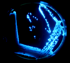
Figure 1. Photo of a Petri plate in the dark, showing the glowing spots from bioluminescent bacteria.
2. The requirement for oxygen. Pass a stream of nitrogen into the Petri dish, keeping the lid in place, but not too firmly that it presses on the rubber tubing and stops the flow of nitrogen. In a few minutes, you’ll see the glow diminish. Allow the air back in, and the glow will return.
3. Culture bioluminescent bacteria. Flame a wire loop, and when cool, use it to pick up a little of one of the single glowing colonies. This should represent a clone from a single bacterium. Inoculate it into the liquid culture using the sterile procedure. Secure the flask on a shaker, and shake so that it aerates for about 24 h, or to the point that the culture is like a soup and glows brightly.
A cell cake is prepared by centrifuging off the liquid. Here you need access to a preparative centrifuge. Resuspend the cake in about 50 mL of distilled water, cool to ice temperature, and sonicate it with about 20 s bursts, recooling to ice temperature in between bursts. This releases the cell proteins, including luciferase, into the liquid, and centrifuge again to remove the cell walls. This time keep the liquid, as this is your crude luciferase preparation.
See also: Isolation of Marine Bioluminescent Bacteria
4. In vitro bioluminescence reaction. To 1 mL of the crude luciferase preparation, add 1 drop of the dodecanal (or decanal) suspension, and 1 drop of FMN solution. Take the test tube, the NADH solution, and a Pasteur pipette into a dark room. Allow about 10 minutes in the dark for your eyes to dark adapt, then add 1 drop of NADH and mix by gentle shaking. The NADH reduces the FMN to FMNH2, an essential substrate for the luciferase, catalyzed by a second enzyme that accompanies the luciferase preparation. The bioluminescence will first be bright, then slowly dim as the reaction uses up the substrates.
SUPPLIES
Chemicals. These can all be purchased from any vendor of laboratory supplies. Glycine H(NH2)COOH (2 g); ammonium bicarbonate NH4HCO3 (1 g); adenosine triphosphate ATP (0.3 g); magnesium chloride MgCl2 (0.5 g); hydrochloric acid HCl (1 N, 10 mL); sodium hydroxide NaOH (1 N, 10 mL); acetic acid CH3COOH (10%, 10 mL); sodium chloride NaCl (10 g); tribasic sodium phosphate Na3PO4 (1 g); dibasic sodium phosphate Na2HPO4 (5 g); dibasic ammonium phosphate (NH4)2HPO4 (1 g); monobasic potassium phosphate KH2PO4 (5 g); magnesium sulfate MgSO4 (1 g); distilled water (2 L); bactopeptone (5 g Difco); bacto-agar (3 g Difco); reduced nicotine adenine dinucleotide NADH (0.3 g); flavin adenine dinucleotide FMN (0.2 g); dodecanal (0.5 mL); nitrogen gas N2 (lecture bottle, regulator, tubing).
Equipment. Mortar and pestle; filter paper and funnel or a table-top centrifuge with 1 mL tubes; test-tubes 10 mL (20); plastic freezer bag (5); pasteur pipettes (25); syringe 1 mL and needle (5) ; pH meter or pH paper; analytical balance and accessories; steam sterilizer; microbiological wire loop (2); petri dishes (25); hot plate (1); bacterial culture 500-mL flask shaker (1); sonic dismembranator or equivalent device for cell disruption (Fisher catalog).
See also:
Sea Fireflies (Ostracods) (PDF)
Dinoflagellates and Red Tides
Edith Widder: Glowing Life in an Underwater World
Scroll down to VIDEO (17 min)
READING LIST
McCapra, F. (2000) Demonstrations of chemiluminescence. Methods Enzymol. 305, 633-659.
Hammer, M., and Andrade, J.D. (2000) Bioluminescence as a classroom tool for scientist volunteers. Methods Enzymol. 305, 660-672.
Ward, W.W., Swiatek, G.C., and Gonzalez, G.D. (2000) Green fluorescent protein in biotechnology education. Methods Enzymol. 305, 672-680.
Kratasyuk V.A., and I. Y. Kudinova, I.Y. (1999) Practical enzymology course based on Bioluminescence. Luminescence 14, 189-192.
10/20/10
01/27/11